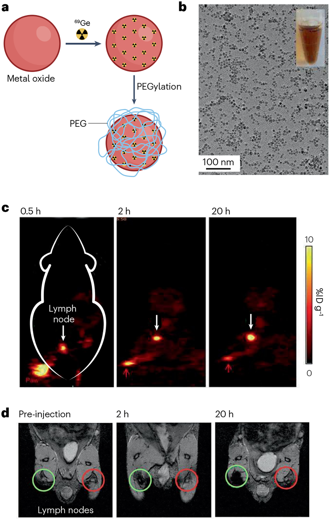
Fig. 5 |. Superparamagnetic iron oxide nanoparticles labelled with 69Ge for lymph node mapping via positron emission tomography and MRI.

a, Schematic illustration of chelator-free synthesis of 69Ge-iron oxide nanoparticles (IONPs). b, Micrograph of IONPs from transmission electron microscopy. c, Lymph node (white arrow) imaging with positron emission tomography after injection of 69Ge-IONPs into left paw (red arrow) of a mouse. d, Lymph node (green circle) mapping with MRI before and after injection of 69Ge-IONPs into left paw of a mouse. Contralateral lymph node is indicated by red circle. %ID g−1, percentage of injected dose per gram of tissue; PEG, polyethylene glycol. Reprinted with permission from ref. 188, Wiley.
