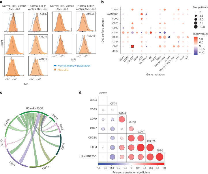
Fig. 2. Cell surface expression of U5 snRNP200 in AML and coexpression patterns with other AML-associated antigens.
a, Histograms of MFI on live HSCs (live Lin−CD34+CD45dimCD90+CD38−) and lympho-primed MPP cells (LMPPs; live Lin−CD34+CD38−CD90−CD45RA+ cells) from normal bone marrow (blue lines) versus on AML leukemia stem cells (LSCs; live CD34+CD38−CD90−CD45RA+ cells) for five distinct patients with AML whose bulk leukemic cells express cell surface U5 snRNP200. b, Bubble plot summarizing surface antigen expression and mutation status patterns. Bubbles with dark circle outlines highlight patterns that reach statistical significance (that is, CD25 expression and DNMT3A mutations co-occur, whereas CD33 and RUNX1 mutations are mutually exclusive). log-transformed P values were determined by one-sided Fisher test, with positive values indicating positive associations and negative values indicating negative associations. c, Circos plot depicting coexpression between U5 snRNP200 and known AML-associated surface antigens on AML patient bone marrow blasts. d, Correlogram depicting Pearson correlation coefficients of surface antigen intensity on AML patient bone marrow blasts.

