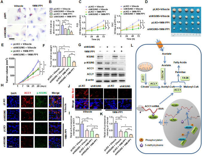
Fig. 8. Blockade of CDK13/NSUN5/ACC1 pathway-mediated fatty acid synthesis inhibits PCa progression.
A C4-2 cells were transfected with shNSUN5 and then treated with or without 1NM-PP1 (10 μM), lipid accumulation was measured by ORO straining. B Quantitative analysis of ORO staining in (A). C PC3 and C4-2 cells were treated as in (A), and cell viability was assessed by MTS assay. D Xenograft tumor models in nude mice were established by implanting PC3 cells with stable knockdown of NSUN5. From the first day, the mice were intraperitoneally injected with 20 mg/kg 1NM-PP1 every two days. Representative tumor sizes in each group were shown. E Tumor volumes were measured with calipers and calculated by the formula: (length × width2)/2. F Wet weight of the xenograft tumors was determined after tumor resection. G Western blotting detected the expression of NSUN5, p-NSUN5, ACC1 and ACLY proteins in xenograft tumors. H The expression of ACC1 and p-NSUN5 in xenograft tumors was detected by double immunofluorescence staining. Red: ACC1; green: p-NSUN5; blue: DAPI. Scale bar = 25 μm. I Quantitative analysis of fluorescence intensity of ACC1in (H). J Nile red staining detected the lipid deposition in xenograft tumors. Red: lipids; Blue: nuclei. Scale bar = 25 μm. K Quantitative analysis of Nile red staining in (J). L A proposed model illustrating the CDK13/NSUN5/ACC1 regulatory pathway-mediated fatty acid synthesis and lipid deposition. All data are expressed as the mean ± SEM of 3 independent experiments. *P < 0.05, **P < 0.01 vs. their corresponding controls.

