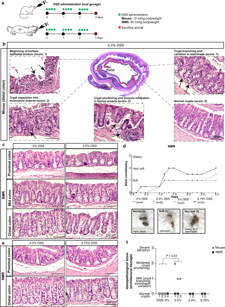
Fig. 6. Histopathological assessment of NMR intestine after dextran sodium sulphate (DSS) treatment.
a Schema illustrating the experimental design for administering DSS via oral gavage at specific time points (green dots) and days of tissue harvest (red dots) in C57BL/6J mice (top) and wild-caught NMRs (bottom). b Centre, representative colonic gut roll of 2.5% DSS-treated mouse, with 30% of distal colon showing damage which is demarcated in red. Magnified images highlighting epithelial surface erosion, crypt damage and inflammation (black arrows) in DSS-treated mice. Undamaged crypts are also shown. c Haematoxylin and eosin stained images of the proximal, mid, and distal colons comparing control and 2.5% DSS treated NMRs. d Top, gradual change over 3 days in the stool consistency of NMRs subjected to DSS treatment at different concentrations [0% (n = 4 animals), 2.5% (n = 6 animals), 5.0% (n = 2 animals) and 8.75% (n = 2 animals)]. Bottom, photographs show three different stool consistencies observed in DSS-treated NMRs (hard, black: Score 0; soft and yellowish: Score 1; very soft, misshaped and slightly watery: Score 2). e Haematoxylin and eosin staining showing no damage in the proximal and distal colons of NMRs treated with 5.0% and 8.75% DSS. f Histopathological scores showing mild to moderate damage in DSS-treated mice (n = 3 animals per group, P-value = 0.03, two-tailed Wilcoxon rank-sum test) while no damage was seen in any of the DSS-treated NMRs (2.5%, 5% and 8.75%; n = 2 animals per treatment group). Scale bars are indicated on the images (100 µm).

