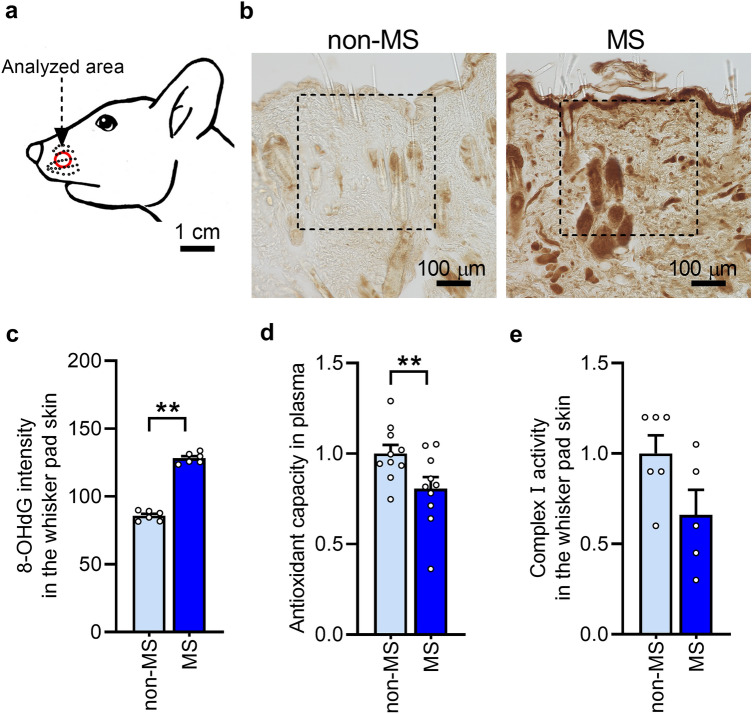
Figure 2.
Oxidative stress in the whisker pad skin following maternal separation (MS). (a) Analyzed area of the whisker pad skin. (b) Microphotograph images of representative 8-hydroxy-deoxyguanosine (8-OHdG) immunoreactivity in the whisker pad skin of non-MS and MS rats at 7 weeks. Dotted square in the images indicate analyzed region. Scale bars indicate 100 µm. (c) 8-OHdG intensity in the whisker pad skin (n = 6 in each). **P < 0.01, unpaired t-test. (d) Plasma antioxidant capacity of MS and non-MS rats (n = 10 in each group). **P < 0.01, unpaired t-test. (e) Complex I activity in the whisker pad skin of MS and non-MS rats (n = 5–6 in each group). P = 0.07, unpaired t-test.

