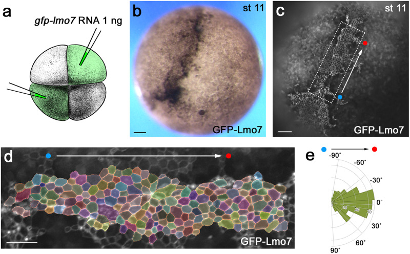
Fig. 4. Lmo7-expressing ectoderm as a model of apical domain heterogeneity.
a Scheme of the experiment. GFP-Lmo7 RNA (1 ng) was injected in two oppositely localized animal blastomeres in four-cell stage embryos. Adapted from Xenopus illustrations © Natalya Zahn (2022), Xenbase (www.xenbase.org RRID:SCR_003280)75. b Representative GFP-Lmo7-expressing embryo at stage 11. Hyperpigmentation was observed in 90–95% embryos (n > 100). c Still image from Movie 1 shows GFP-Lmo7 fluorescence from the embryo in (b). d Segmented image of the rectangular area in (c). e Representative rose plot from n = 792 cells from one embryo depicts cell orientation with respect to the injection axis (white arrow connecting red and blue dots in c–e). The data represent at least three experiments. Scale bars are 100 μm in (b, c) and 50 μm in (d).

