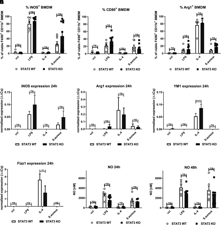
FIGURE 2.
No changes in classical M1/M2 markers and NO induction in STAT3-deficient compared with WT BMDM upon S. aureus stimulation. STAT3 WT and KO BMDM were stimulated with vcl, LPS (100 ng/ml), IL-4 (20 ng/ml), or S. aureus (MOI 10) for a total of 24 or 48 h following addition of penicillin-streptomycin at 2 h of stimulation. Supernatants were harvested, and cells were further processed for downstream analysis. (A–C) For flow cytometric analysis of BMDM, live/dead staining, as well as surface marker staining (CD80), followed by permeabilization and intracellular staining (iNOS, Arg1), was performed at 24 h. Gating was performed on single viable F4/80+ CD11b+ BMDM. Percentage of (A) iNOS+, (B) CD80+, and (C) Arg1+ F4/80+ CD11b+ BMDM. (D–G) In selected experiments, cells were processed for RNA extraction by the TRIzol method. Following cDNA generation from RNA, qPCR of (D) iNOS, (E) Arg1, (F) YM1, and (G) Fizz1 was run. Pooled data (mean with SEM) from n ≥ 3 different experiments are shown. (H and I) NO measurement in supernatants from STAT3 WT and KO BMDM stimulated with vcl, LPS, IL-4, or S. aureus for (H) 24 and (I) 48 h. Pooled data (mean with SEM) from n ≥ 5 different experiments are shown. Mixed-effects model with Bonferroni analysis for multiple comparisons was used for statistical analysis. p > 0.05 not significant; **p ≤ 0.01, ****p ≤ 0.0001.

