FIGURE 1.
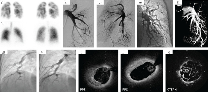
Comparative representative images of peripheral pulmonary artery stenosis (PPS) and chronic thromboembolic pulmonary hypertension (CTEPH). a, b) In a patient with PPS, a) lung perfusion scintigraphy showed multiple wedge-shaped defects and b) lung ventilation scintigraphy showed normal findings. c–e) Pulmonary angiography (PAG) from typical PPS cases presenting with c, d) multiple stenosis and e) tortuosity in segmental pulmonary arteries. f) Three-dimensional image constructed from PAG showed pulmonary arterial stenosis. g, h) PAG images g) before and h) after percutaneous pulmonary angioplasty. i–k) Optical coherence tomography showed thickening of the medial layer of the pulmonary artery and the vascular characteristics were different from those of CTEPH. These images were obtained in a few cases.

