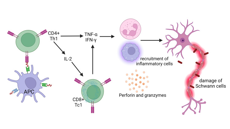
Figure 5. The mechanism of nerve damage in type 1 leprosy reaction.
Activated macrophages release pro-inflammatory cytokines, such as tumor necrosis factor-alpha (TNF-α), interleukin-1 beta (IL-1β), and interleukin-6 (IL-6). The cytokines caused increasing endothelial barrier permeability, thereby promoting immune cell transmigration towards the inflammatory locus. As a result, this series of physiological processes ultimately manifests localized edema, erythema, and increased temperature. Furthermore, it is essential to highlight that macrophages can release lysosomal enzymes, complement components, and reactive oxygen species in a localized anatomical area. The complex mechanism has a crucial role in the progression of tissue damage. Perforin facilitates pore formation within the target cell's membrane and promotes the entry of granzymes into the cellular environment, thus initiating the complex series of apoptosis.
APC: antigen-presenting cell, IFN‐γ: interferon‐gamma, IL: interleukin
This figure was created by NA, one of the authors of this article, with Biorender.com.

