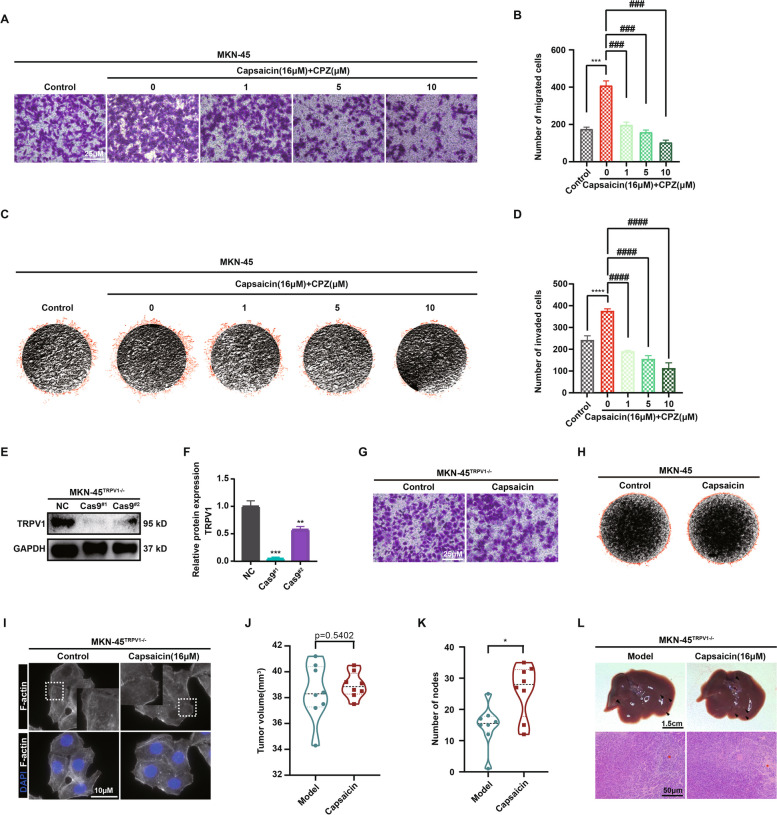
Fig. 4.
Capsaicin retarding the progression of GC was partially mediated through the functional TRPV1. Representative images (A) and quantitative analysis (B) for the migration of MKN-45 cells in the absence and presence of capsazepine (CPZ) using the transwell migration assay. Visual fields were selected randomly from each sample (scale bars: 250 μm). Representative images (C) and quantitative analysis (D) for the invasion of MKN-45 cells invasion in the absence and presence of CPZ using the 3D-invasion system (n = 3). Representative images (E) and quantitative data (F) for the protein level of TRPV1 in the MKN-45 cells and MKN-45TRPV1−/− cells (n = 3). GAPDH was used as a loading control. G Representative images of migration of MKN-45 and MKN-45TRPV1−/− cells using the transwell migration assay. Visual fields were selected randomly from each sample (scale bars: 250 μm). H Representative images of invasion of MKN-45 and MKN-45TRPV1−/− cells using the 3D-invasion system. I Representative images of fluorescence staining for the actin cytoskeleton in the MKN-45TRPV1−/− cells after the treatment of 16 μM capsaicin for 24 h (scale bar: 10 μm). The tumor volume (J) and the number of liver metastatic nodules (K) of MKN-45.TRPV1−/− orthotropic tumors in the normal diet and 100 mg/kg capsaicin diet groups were shown. L Representative images for the liver (upper panel) and H&E staining sections (bottom panel) (scale bars: 1.5 cm for liver images and 50 μm for H&E-stained images)

