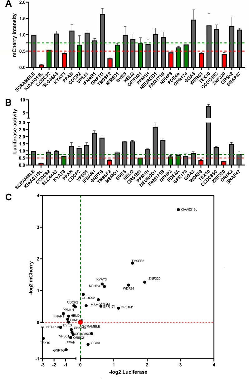
Fig 2.
rAAV2.5T transduction in the 28 top-ranking gene-silenced HeLa cells. (A and B) Relative expression of luciferase and mCherry in gene-silenced cells. HeLa cells seeded in 24-well plates were transduced with a shRNA-expressing lentiviral vector. After selection in puromycin, the cells were transduced with rAAV2.5T at an MOI of 20,000 DRPs/cell. At 3 days post-transduction, mCherry expression was imaged under a fluorescence imager (A), and the luciferase activity was measured using a firefly luciferase detection reagent (B). All data had three repeats and were normalized to the non-target (NT) control. The green and red dashed lines indicate 75% and 50% of the NT control, respectively. Data shown are means with a standard deviation (SD) from three replicates. (C) Ranks of the candidates in quadrant I. The x-axis shows −log2 luciferase intensity, and the y-axis shows −log2 mCherry intensity. All the cells silenced with gene names shown in quadrant I had decreases in both luciferase and mCherry expressions.

