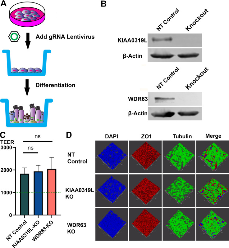
Fig 4.
Generation of gene knockouts in HAE-ALI cultures (A) Knockouts of KIAA0319L and WDR63 in HAE-ALI cultures. A schematic diagram shows the generation of gene knockouts of CuFi-8-derived HAE-ALI cultures. CuFi-8 cells were cultured in collagen-coated 100-mm dishes until confluent, and then gRNA-expressing lentiviruses were added to the dishes. Under the selection of puromycin, the CuFi-8 cells were single-clone expanded for the production of a gene knockout cell line. Expanded gene knockout cells were transferred onto the transwell for polarization at ALI. (B) Gene knockdown efficiency. Western blotting and genomic DNA sequencing validated the knockouts of KIAA0319L and WDR63 in HAE-ALI cultures. Cells harvested from the transwells were extracted for proteins and DNA, respectively. Western blotting detected both the non-target control and knockout groups, KIAA0319L and WDR63, respectively. β-Actin was detected as a loading control. The genomic DNA was amplified for sequencing the mutations at the gRNA targeting sequences of the KIAA0319L and WDR63 genes, respectively. The gene sequences show the original gene sequence, the non-target gene sequence, and the gene knockout sequence. (C) TEER measurement. After 1 month of differentiation at ALI, HAE-ALI cultures, non-target NT (NT) control, KIAA0319L-KO, and WDR63-KO, were detected for TEER values. Data shown are means with an SD of three replicates. (D) Three-dimensional confocal imaging of ZO-1 and β-tubulin expression in gene knockout HAE-ALI. NT, KIAA0319L-KO, and WDR63-KO HAE-ALI cultures were fixed and co-stained with anti-β-tubulin IV (green) and anti-ZO-1 (red) antibodies. Nuclei were stained with DAPI (blue). A set of confocal images was taken at a magnification of ×40 (Leica SP8 STED) from the stained piece of the epithelium from the objective (z-axis) and reconstituted as a three-dimensional image as shown in each channel of fluorescence.

