Fig 5.
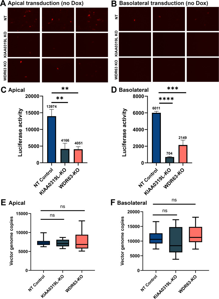
Knockouts of KIAA0319L and WDR63 cause a significant decrease in the transduction efficiency of rAAV2.5T in HAE-ALI cultures but not vector internalization. HAE-ALI cultures were transduced with rAAV2.5T at an MOI of 20,000 DRPs/cell from the apical (left panels A, C, and E) and basolateral chambers (right panels B, D, and F), respectively. At 16 hours post-transduction, both the apical and basolateral chambers were refreshed with culture media. (A and B) mCherry expression. Images were taken under a fluorescence imager at 5 days post-transduction. (C and D) Luciferase activity assay. Luciferase activity was measured at 5 days post-transduction. Data shown are means with an SD from three replicates. (E and F) Vector internalization assay. HAE-ALI cultures were incubated with rAAV2.5T at an MOI of 20,000 DRPs/cell from the apical and basolateral chambers, respectively, for 2 hours at 37°C. Then, the viruses were removed, and both chambers were treated with Accutase three times. Viral DNA was extracted using the pathogen viral DNA collection kit (Zymo) and quantified via quantitative polymerase chain reaction (qPCR) using an mCherry gene-targeting probe to detect the entered viral genomes. The boundary of the box closest to zero indicates the 25th percentile, a black line within the box marks the median, and the boundary of the box farthest from zero indicates the 75th percentile. Whiskers above and below the box indicate the 10th and 90th percentiles.

