Fig 7.
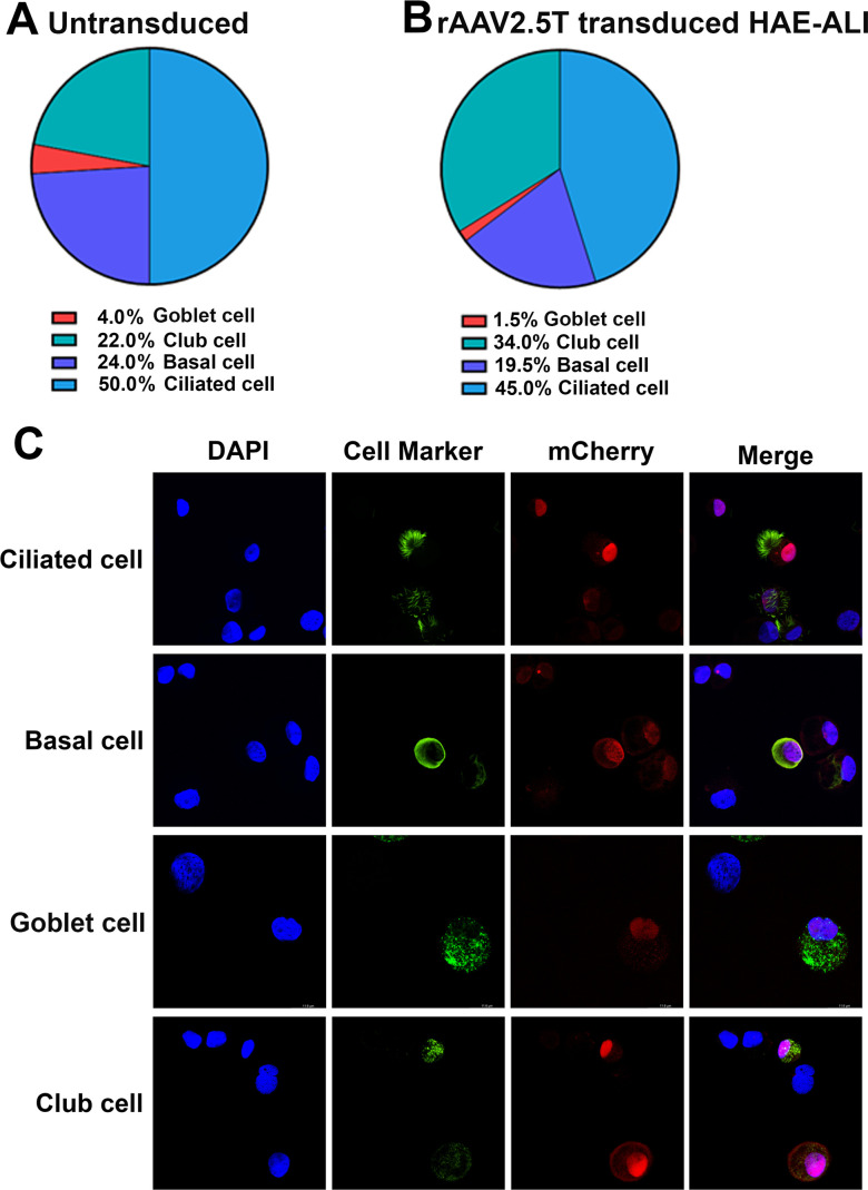
Analysis of rAAV2.5T-transduced cell types in HAE-ALI cultures. HAE-ALI cultures were transduced with rAAV2.5T at an MOI of 20,000 DRPs/cell from the apical chamber or mock transduced. Dox was added to rAAV2.5T at 2.0 µM. (A) Flow cytometry of airway cell types in HAE-ALI cultures. The cells of the mock-transduced HAE-ALI cultures were digested off the transwell membranes, fixed, and permeabilized, followed by immunostaining with the primary antibody against each cell marker and an Alexa 488-conjugated secondary antibody. The stained cells were subjected to flow cytometry, and the percentage of each cell type was calculated. (B) Flow cytometry of airway cells transduced with rAAV2.5T. At 7 days post-transduction, the cells of the rAAV2.5T-transduced HAE-ALI cultures were digested off the transwell membranes and sorted on a BD FACSAria III Cell Sorter. mCherry-positive cells were sorted by FACS, fixed, and permeabilized, followed by immunostaining with the primary antibody against each cell marker and an Alexa 488-conjugated secondary antibody. The stained cells were subjected to flow cytometry, and the percentage of each cell type was calculated. (C) Immunofluorescence assays. At 7 days post-transduction, the cells of rAAV2.5T-transduced HAE-ALI cultures were digested off the transwell membranes, cytospun to slides, fixed, and permeabilized, followed by immunostaining with an antibody against each cell marker. The stained cells were subjected to imaging under confocal microscopy at a magnification of ×100 (CSU-W1 SoRa, Nikon).

