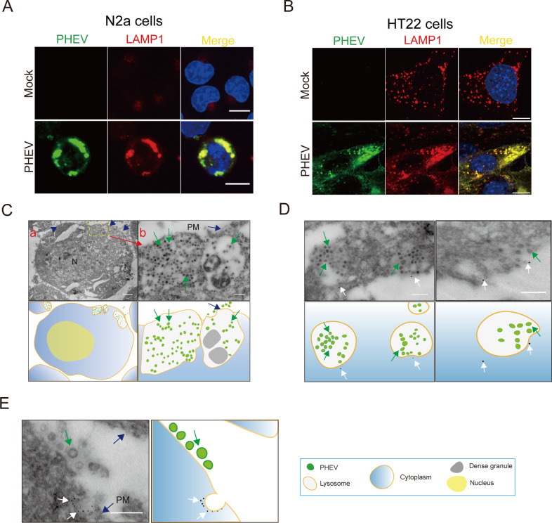
Fig 1.
PHEV is enriched in late endosomes/lysosomes during replication. (A and B) Colocalization of PHEV and LAMP1 in N2a and HT22 cells, respectively. Mock- and PHEV-infected cells were immunostained with anti-LAMP1 (red) and anti-PHEV (green) antibodies at 48 hpi. Scale bar, 10 µm. (C) TEM images of PHEV-infected N2a cells at 48 hpi. Blue arrows: The plasma membrane; Green arrows: PHEV. Scale bar, 1 µm (A) and 200 nm (B). (D and E) PHEV-infected N2a cells at 48 hpi were processed for immuno-EM and labeled with anti-LAMP1 and 12 nm colloidal gold. Blue arrows: the plasma membrane; White arrows: the colloidal gold-labeled LAMP1; Green arrows: PHEV. Scale bar, 200 nm. Representative images are shown.

