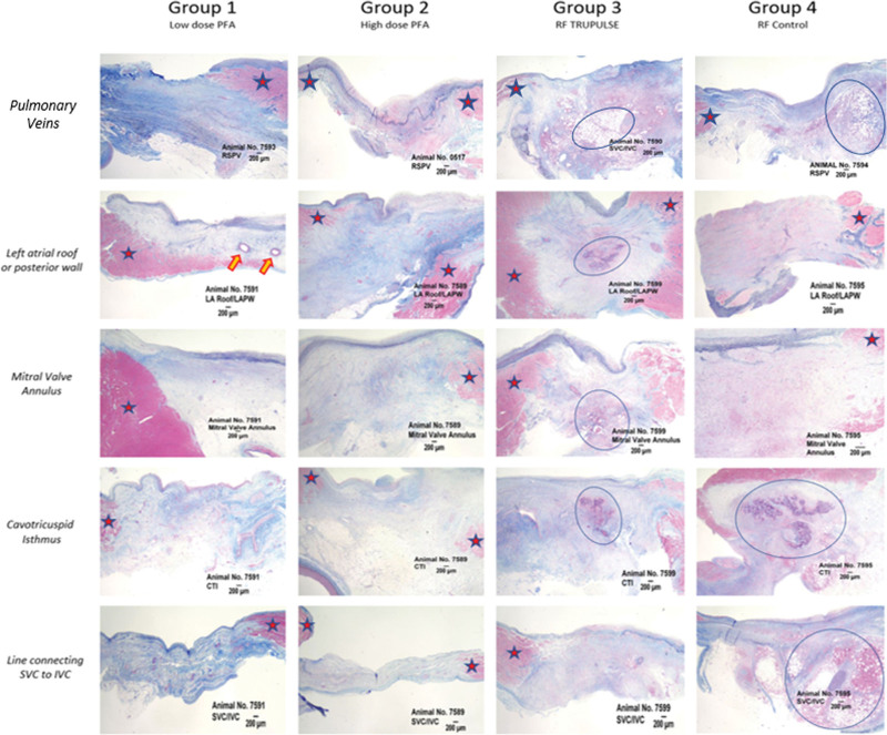
Figure 4.
Histological architecture of the evaluated lesions across different anatomic locations and different study arms. Pulsed field ablation (PFA) vs radiofrequency (RF) lesions at the investigated ablated sites (pulmonary veins, left atrial roof or posterior wall, mitral valve annulus or mitral isthmus, cavotricuspid isthmus [CTI], and, finally, the posterior right atrium through a line connecting the superior vena cava [SVC] to the inferior vena cava [IVC]). Greater inflammatory response and necrotic burden were observed in animals treated with RF vs PFA. Red stars represent healthy tissue and blue circles areas of inflammation and necrosis. Orange arrows showing intact mid-myocardial vessels in PFA lesions. LA indicates left atrium; PW, posterior wall; and RSPV, right superior pulmonary vein.

