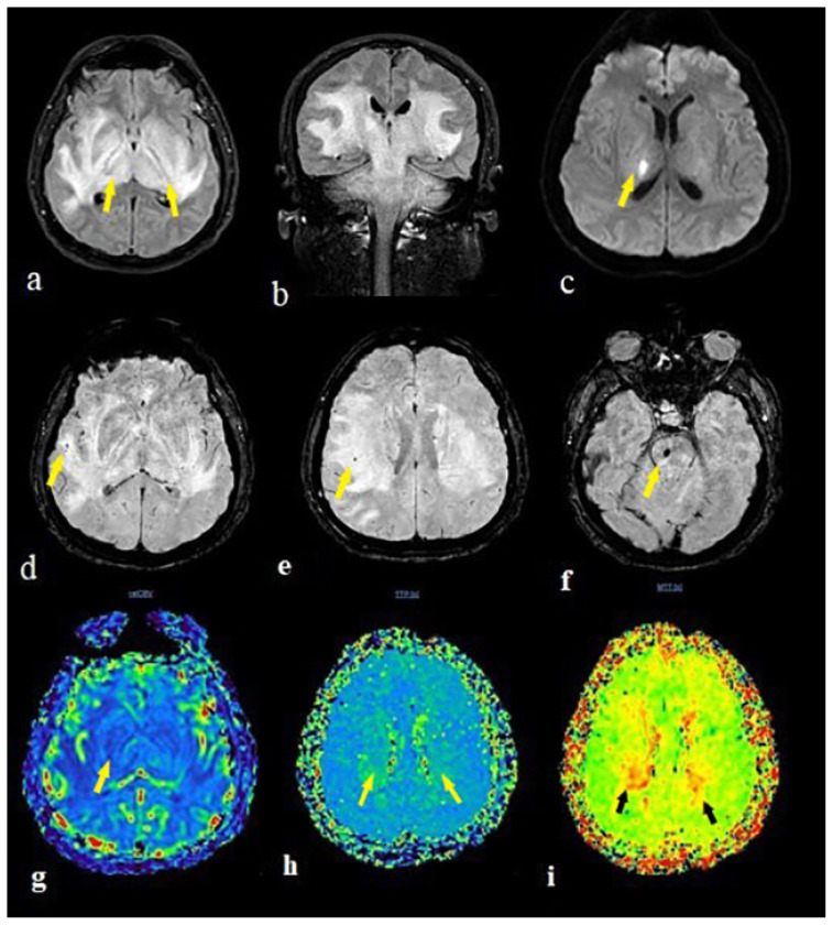
Figure 4.
The brain MRI of a 41 years old male patient with encephalitis showed extensive hypertens signal in bilateral cerebral hemisphere white matter, thalamus, basal ganglia corticospinal tract, brain stem and serebellum on FLAIR images (a,b). The diffusion restrction occured only in the right thalamus on DWI sequence (c). There were microhemorrhages in the pons(f) and right parietal deep white matter (d,e) on SWI images. The perfusion MR imaging showed any CBV increment (g) with prolongation in bilateral thalamus and right temporal lobe on time to peak (h) and mean transit time (i) maps (arrows).

