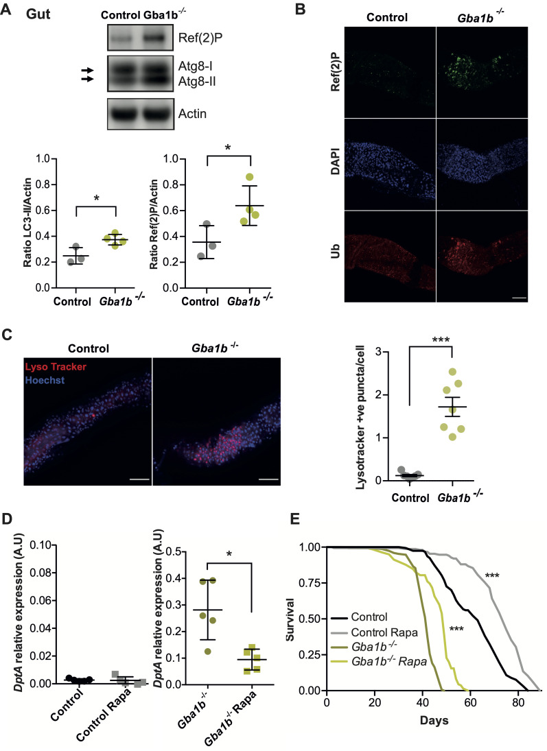
Fig 7. Loss of Gba1b results in gut autophagy impairment and administration of rapamycin rescues Gba1b-/- immune phenotypes.
(A) Gba1b-/- fly guts show significant accumulation of Atg8a-II and Ref(2)P proteins relative to control guts (*p = 0.024 and *p = 0.049, respectively). Unpaired t-tests; data are presented as mean ± SD (n = 3–4). (B) Immunostainings of guts labelled for Ref(2)P (green), DAPI (blue) and ubiquitin (Ub, red). Gba1b-/- flies display a higher number of aggregates of Ref(2)P and ubiquitinated proteins in the midgut. Scale bar 100 μm. (C) Gut staining with LysoTracker (red) and Hoechst (blue) of 3-week-old flies reveals an increased number of LysoTracker puncta in Gba1b-/- flies (***p<0.0001; unpaired t-test). Scale bar 50 μm. (D) DptA transcript levels are reduced on qRT-PCR analysis of the guts of 3-week-old Gba1b-/- flies treated with Rapamycin (Rapa) compared to non-treated flies (*p = 0.0159). No significant differences are found in control flies (ns = 0.5317). Mann Whitney tests; data are presented as mean ± SD (n = 5 per condition). (E) The survival of Rapa treated control and Gba1b-/- flies (n = 150) is significantly increased. Log-rank tests: Gba1b-/- vs Gba1b-/- Rapa, *** p<0.0001; Control vs Control Rapa *** p<0.0001.

