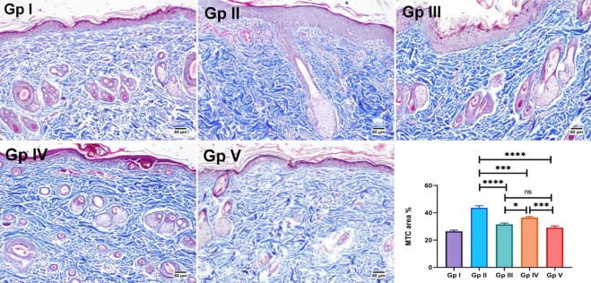
Fig 7. MTC-skin-stained sections for evaluation of collagen fibers.
Gp I (normal control group) showed normal collagen bundles. Abnormal accumulation of collagen fibers is found in Gp II (UVB group) and Gp IV (FO group). Apparently normal collagen is shown in Gp V (FO-SLNs group). Chart present the area % of MTC staining in different groups. Data are presented as mean ±SE. Significant difference was considered at p<0.05.

