
FIGURE 3.
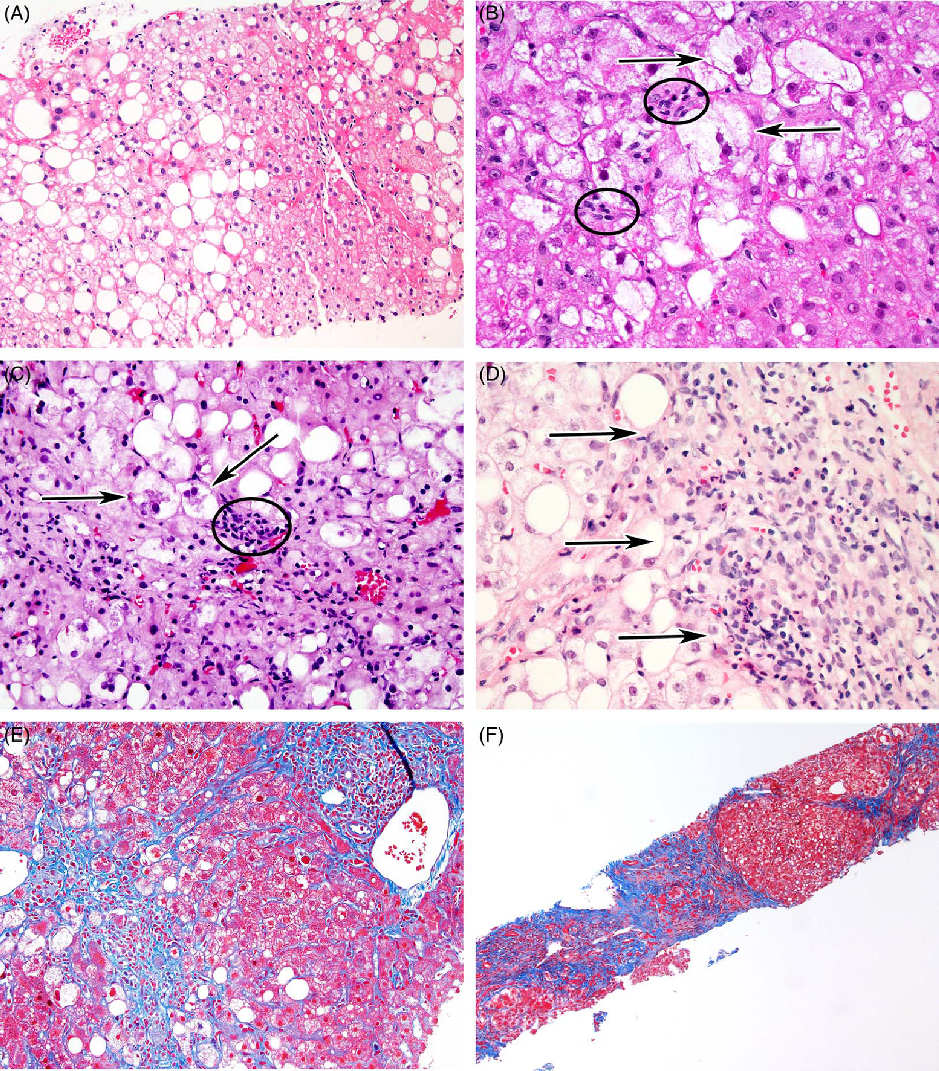
Histology of NAFLD. Liver biopsy shows characteristic features of the spectrum of NAFLD. (A) Hepatic steatosis (typically zone 3) without ballooned hepatocytes or fibrosis [hematoxylin and eosin (H&E), ×200]. (B) Multiple ballooned hepatocytes with Mallory-Denk bodies (arrows) and mild lobular inflammation (circles) (H&E, ×400). (C) Ballooned hepatocytes (arrows) with moderate lobular inflammation (circle) (H&E, ×200). (D) Some cases of steatohepatitis may show significant portal inflammation and interface hepatitis (arrows) (H&E, ×200). (E) Dense perisinusoidal and periportal fibrosis (blue stain), with a thin connecting fibrotic bridge (Masson trichrome, ×200). (F) Cirrhosis (nodule formation) due to steatohepatitis (Masson trichrome, ×100).