. 2023 Dec 21;12:RP92769. doi: 10.7554/eLife.92769

Figure 4. Cfap52 deficiency in mice leads to male sterility, with spermatozoa showing defective head-tail connections and flagella.

(A) Schematic illustration of the targeting strategy for generating Cfap52-KO mice by using CRISPR/Cas9 technology. A detailed procedure is described in the Materials and methods. (B) Representative results of PCR-based genotyping using mouse tail DNA. (C) Immunoblotting of CFAP52 was performed in the testis protein lysates of WT mice and Cfap52-KO mice. β-actin served as a loading control. Western blotting experiments were repeated three times with similar results. (D, E) Fertility assessment experiments were performed on three adult Cfap52-KO male mice and three WT male littermates for 2 months. Of the 20 female mice mated with Cfap52-KO male mice, no pregnancy was observed. (F, G) Sperm counts were counted with a fertility counting chamber under a light microscope, and total motility was assessed by a computer-assisted sperm analysis (CASA) system. Data are presented as the mean ± SEM (n=3 each group), Student’s t test, *p<0.05; ***p<0.001. (H) Morphological analyses of spermatozoa in WT mice and Cfap52-KO mice by Papanicolaou staining. First row, normal morphology of spermatozoa from WT mice; second row, headless spermatozoa in Cfap52-KO mice; third row, spermatozoa with defective head-tail connection in Cfap52-KO mice; fourth row, short-tailed spermatozoa in Cfap52-KO mice. The arrowheads indicate the ‘pinhead’, and the arrows indicate the defective head-tail connection. Scale bars, 5 μm. (I) Percentage of spermatozoa with normal morphology and each type of defect in WT mice and Cfap52-KO mice. At least 100 spermatozoa were counted for each mouse. Data are presented as the mean ± SEM (n=3 each group), Student’s t test, **p<0.01; ***p<0.001.

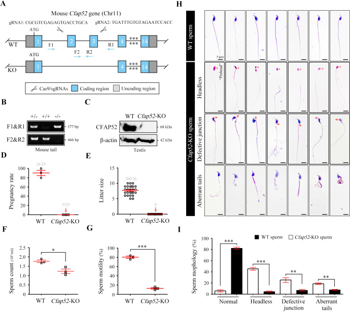
Figure 4—source data 1. Primers for Cfap52-KO mouse genotyping.
Figure 4—source data 2. Original blots of Figure 4C.

Figure 4.

Figure 4—figure supplement 1. Animal report of generation of Cfap52-KO mice.

Figure 4—figure supplement 1.

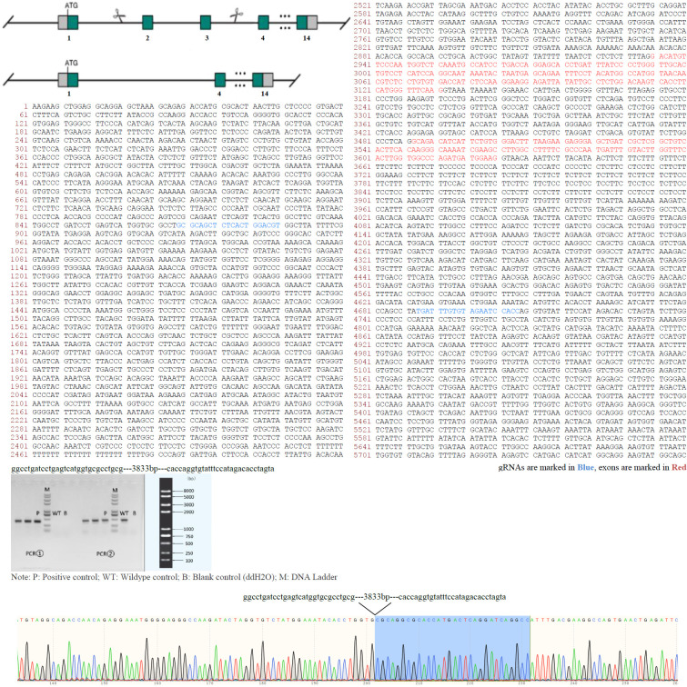
Genomic region of mouse Cfap52 locus. The mouse Cfap52-201 transcript (ENSMUST00000021287.11) is located on mouse chromosome 11. Fourteen exons have been identified and exons 2~3 were selected as the knockout region. gRNAs are marked in blue or underlined and exons are marked in red. Cas9 mRNA and gRNAs were co-injected into fertilized eggs for KO mouse production. The pups will be genotyped by PCR followed by sequence analysis. F0 found animals were bred to wildtype mice to test germline transmission and F1 animal generation. Positive F1 animals were confirmed by PCR and Sanger sequencing (~3833 bp deletion containing exons 2~3 of Cfap52). Sequence of gRNAs, gel image, and Sanger sequencing were shown.
Figure 4—figure supplement 2. Cfap52-KO mice develop hydrocephalus.

Figure 4—figure supplement 2.

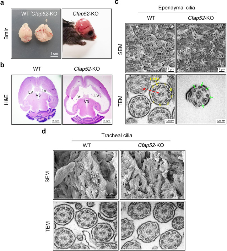
(a) Enlarged brain hemispheres and hydrocephalus of Cfap52-KO mice. Scale bar, 1 cm. (b) Histological analysis of brain sections revealed enlarged lateral ventricles (LV) in 3-week-old Cfap52-KO mice. Scale bars, 2 mm. (c) Scanning electron microscope (SEM) images of the ependymal cilia from WT and Cfap52-KO brains, showing the sparse ependymal cilia in Cfap52-KO mice. Scale bars, 2 μm. Transmission electron microscope (TEM) images of ependymal cilia from WT and Cfap52-KO brains, showing the disrupted axonemal structures. Green arrows indicate a partial lack of DMT. CP, central pair; DMTs, doublet microtubules. Scale bars, 100 nm. (d) Scanning electron microscope (SEM) and transmission electron microscope (TEM) images of tracheal cilia from WT and Cfap52-KO mice, showing generally normal morphology and axonemal structure. Scale bars in SEM, 5 μm; Scale bars in TEM, 100 nm. All experiments were repeated three times with similar results.
Figure 4—figure supplement 3. Detailed analysis of infertility in Cfap52-KO male and female mice.

Figure 4—figure supplement 3.

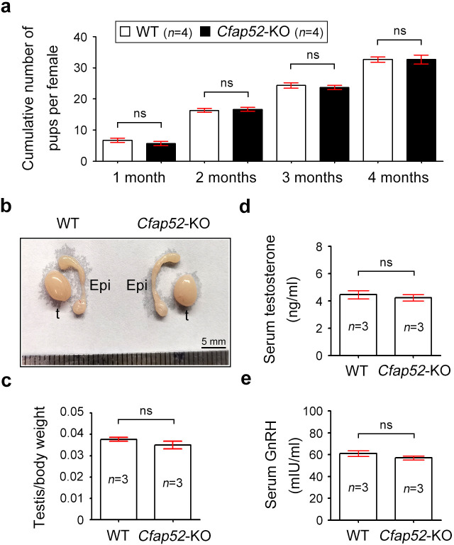
(a) Continuous breeding assay was performed with Cfap52-KO female mice and WT female mice (n=4 each). Female mice were crossed with WT male mice and the average cumulative number of pups per female was shown. Data are presented as the mean ± SEM (three biological repeats), student’s t-test; ns, not significance. (b) Representative reproductive system (testis and epididymis) of WT and Cfap52-KO mice, exhibiting similar morphology and size. Epi, epididymis; t, testis. Scale bar, 5 mm. (c) The testis/body weight ratio of WT and Cfap52-KO mice (n=3 each). Data are presented as the mean ± SEM, student’s t-test; ns, not significance. (d) Elisa measurement of serum testosterone by an ELISA kit (Beyotime, Shanghai, China). (e) The content of GnRH in serum was detected by an ELISA kit (Elabscience, Wuhan, China). Data are presented as the mean ± SEM (three biological repeats), student’s t-test; ns, not significance.
Figure 4—figure supplement 4. Periodic acid-Schiff staining of WT and Cfap52-KO testis sections.

Figure 4—figure supplement 4.

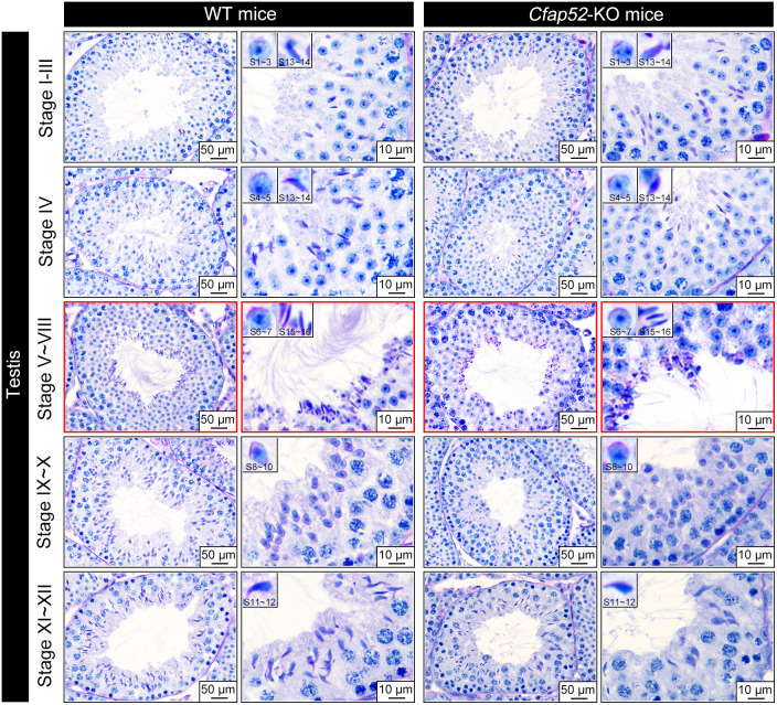
Examination of seminiferous epithelium (stage I~XII) within testes revealed no difference in the population and differentiation of spermatogonia, spermatocyte and round spermatids between WT and Cfap52-KO mice. Acrosome biogenesis of spermatids was also normal in Cfap52-KO mice. An obvious defect was observed at stage V-VIII, showing abnormalities in spermatozoa flagella. Periodic acid-Schiff staining experiments were repeated three times with similar results. Scale bars, 50 μm or 10 μm.