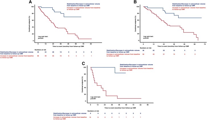
Figure 4.
(A–C) Kaplan–Meier curves for cardiac amyloidosis cohorts stratified according to change in extracellular volume. Patients with increasing extracellular volume (ECV) from baseline to follow-up cardiac magnetic resonance (CMR) imaging had significantly shorter event-free survival (panel A, total cohort; panel B, transthyretin cohort; and panel C, light chain cohort; P for all <0.001). Start date for the follow-up period is the date of follow-up CMR.

