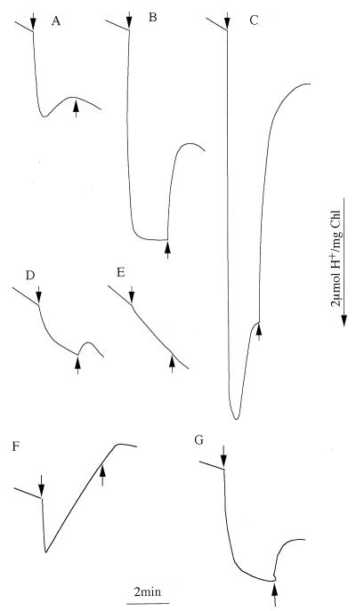
FIG. 2.
Net proton movement in the suspensions of psaAB (A to C), psbDIC/psbDII (D and E), and coxAB (F and G) cells upon switching the light on (arrow down) and off (arrow up). The cells were suspended in 0.2 mM TES-KOH buffer containing 15 mM KCl (A) and NaCl (B to G). DMBQ was added prior to illumination in panels C, E, and F. The chlorophyll concentration in the cell suspension was 1.4 μg/ml for the psaAB mutant and 14 μg/ml for the psbDIC/psbDII and coxAB mutants.

