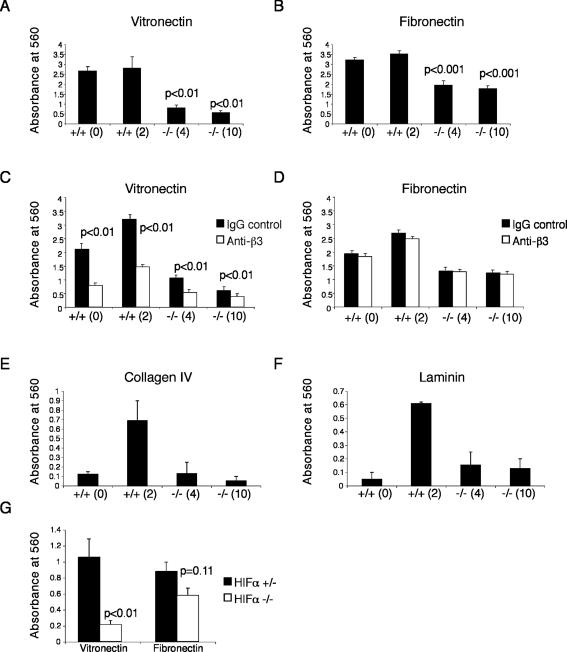
Figure 3.
Arnt-/- TS cells adhere poorly to both vitronectin and fibronectin. Arnt+/+ and Arnt-/- TS cells were plated on strips coated with vitronectin (A) or fibronectin (B) and stained with crystal violet. Solubilized crystal violet incorporated by adherent cells was measured on a plate reader at 560 nm. Two independent wild-type and mutant lines were used. Treatment of TS cells with a blocking antibody for β3 integrin reduces vitronectin (C) but not fibronectin (D). Arnt-/- TS cells adhere to collagen IV (E) and laminin (F) as well as Arnt+/+ TS line 0. Hifα-/- TS cells demonstrate a substantial reduction in adhesion to vitronectin (p < 0.01) and are somewhat reduced in adhesion to fibronectin coated-strips compared with Hifα+/- TS cells (G). Each experiment was performed three times in triplicate. Error bars represent ± SEM. Student's t tests were used for statistical significance.

