Figure 7.
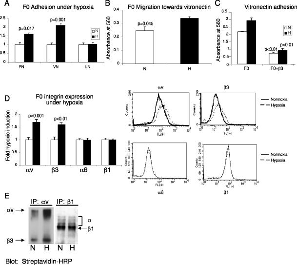
Adhesion to vitronectin and migration toward vitronectin is increased in B16F0 melanoma cells cultured in 1.5% O2 versus 20% O2, via increased cell surface expression of αvβ3. (A) An increase in adhesion to FN and VN is observed in hypoxic B16F0 cells. Adhesion to LN is not changed by hypoxia. (B) Migration toward vitronectin is also enhanced by hypoxia in F0 cells. (C) Adhesion to vitronectin under normoxia and hypoxia is mediated by the β3 integrin because a blocking antibody for β3 integrin diminishes adhesion to vitronectin. (D) B16F0 cells exhibit increased αvβ3 integrin surface expression when treated with 1.5%O2 for 24 h. Surface levels of α6 and β1 integrins are unaltered by hypoxia. FACS analysis for integrins αv, β3, α6, and β1 was performed on cells cultured in either 20% O2 (solid line) or 1.5%O2 (dotted line). (E) B16F0 cells were surface labeled with biotin and immunoprecipitated for αv integrin or β1 integrin. IPs were separated by nonreducing SDS-PAGE and blotted with streptavidin-HRP. Positions of αv integrin, β3 integrin, and β1 integrin are indicated. Error bars represent ± SEM. Student's t tests were performed to determine statistical significance.

