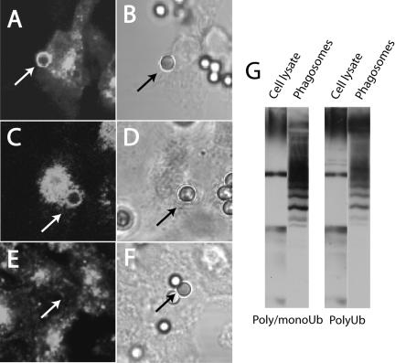
Figure 6.
Ubiquitin is recruited to the phagosome during maturation. (A and B) CHO cells stably transfected with FcγRIIA-GFP were allowed to internalize opsonized latex particles for 20 min. The cells were fixed and permeabilized and immunostained with an antibody that recognizes both poly- and mono-ubiquitin (A). Corresponding DIC image (B). (C–F) Ts20 cells stably transfected with FcγRIIA-GFP were either maintained at 34°C (C and D) or were preincubated at 42°C for 2 h (E and F) before phagocytosis and immunostaining as above. (C–E) ubiquitin immunostaining; (D–F) corresponding DIC images. Arrows point to phagosomes. (G) RAW macrophages were allowed to ingest opsonized latex beads for 12 min at 37°C before homogenization and isolation of phagosomes by sucrose density centrifugation. Purified phagosomes were immunoblotted with an antibody that reacts with both poly- and mono-ubiquitin and another one for poly-ubiquitin alone. An equivalent amount of protein from the starting whole-cell lysate was used as reference. Blots and images are representative of at least three similar experiments of each type.

