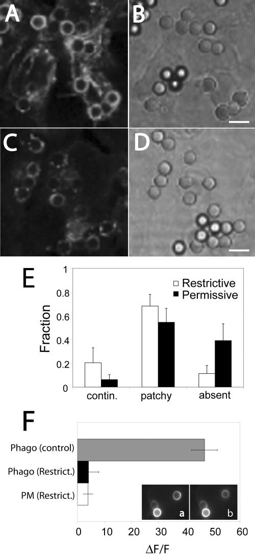
Figure 7.
Impairing ubiquitination prevents the loss of FcγRIIA from the phagosome. Ts20 cells stably transfected with FcγRIIA-GFP were either maintained at the permissive temperature (34°C; C and D) or were preincubated at a restrictive temperature (42°C) for 2 h (A and B) before phagocytosis of opsonized beads for 40 min at 37°C. The cells were then fixed and the distribution of FcγRIIA-GFP analyzed by laser confocal microscopy (A and C). Corresponding DIC images appear in B and D. (E) The fluorescence pattern of each phagosome was categorized as being continuous around the phagosome, patchy (discontinuous) or absent from the phagosome. The fraction of each type is shown in E for both conditions. (F) Quantitation of fluorescence change induced by addition of NH4Cl in the immediate vicinity of the phagosome (Phago) and at the plasma membrane (PM). Ts20 cells stably transfected with FcγRIIA-GFP were incubated at the permissive (control) or restrictive (Restrict.) temperature before ingestion of opsonized latex beads for 45 min at 37°C. Fluorescence emission was acquired before (a) and after the addition of 10 mM NH4Cl (b). Results are presented as the fluorescence change (ΔF) induced by NH4Cl normalized to the total initial fluorescence of the phagosome (F). Data are means ± SE of at least 40 individual determinations from two separate experiments. Size bar, 5 μm.

