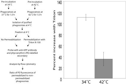
Figure 8.
Measurement of accessibility of the cytosolic tail of FcγRIIA-GFP in isolated phagosomes using flow cytometry. Left panel: flow diagram of the experimental protocol. Ts20 cells stably transfected with FcγRIIA-GFP were incubated at 34 or 42°C for 2 h before ingesting opsonized latex beads. After 1.5 h of phagocytosis, phagosomes were isolated by sucrose density centrifugation, fixed, and either permeabilized with Triton X-100 or left intact. The accessibility of the GFP moiety of the construct, an indicator of the cytosolic tail, was then probed with rabbit anti-GFP and phycoerythrin (PE)-conjugated goat anti-rabbit antibodies. Quantitation was by flow cytometry. Right panel: results are presented as the percentage increase in PE fluorescence induced by permeabilization with Triton. The open bar represents phagosomes from cells at 34°C, and the gray bar represents phagosomes from cells preincubated at 42°C.

