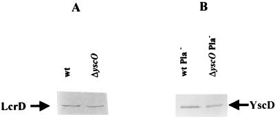
FIG. 5.
Detection of LcrD and YscD in ΔyscO Y. pestis. Y. pestis strains were grown in TMH at 37°C in the absence of Ca2+. The proteins in membrane fractions were separated by SDS-PAGE (12% [wt/vol] acrylamide), transferred to Immobilon P, and analyzed by immunoanalysis with antibodies specific to LcrD or YscD. (A) Y. pestis KIM5-3001 (wt) and Y. pestis KIM5-3001.16 (ΔyscO) were analyzed with anti-LcrD. (B) Y. pestis KIM8-3002 (wt Pla−) and Y. pestis KIM8-3002.3 (ΔyscO Pla−) were analyzed with anti-YscD. The secondary antibody used was conjugated to alkaline phosphatase. Arrows indicate each protein.

