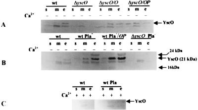
FIG. 6.
Localization of YscO by immunoblot analysis. Y. pestis KIM5-3001 (wt), KIM5-3001.16 (ΔyscO), KIM8-3002 (wt Pla−), and KIM8-3002.3 (ΔyscO Pla−) are shown. Strains carrying plasmids are denoted /O or /OP for pYscO.2 or pYscOP.2, respectively. Bacteria were grown at 37°C in TMH with (+) or without (−) Ca2+ for 5 h prior to harvest. Proteins from bacterial fractions were separated by SDS-PAGE (15% [wt/vol] acrylamide). Antibody raised against a GST-YscO fusion protein was used to detect YscO in soluble (s), total membrane (m), whole-cell (wc), or culture medium (e) proteins. All panels were analyzed with alkaline phosphatase. Arrows indicate the various proteins that were potential candidates for YscO.

