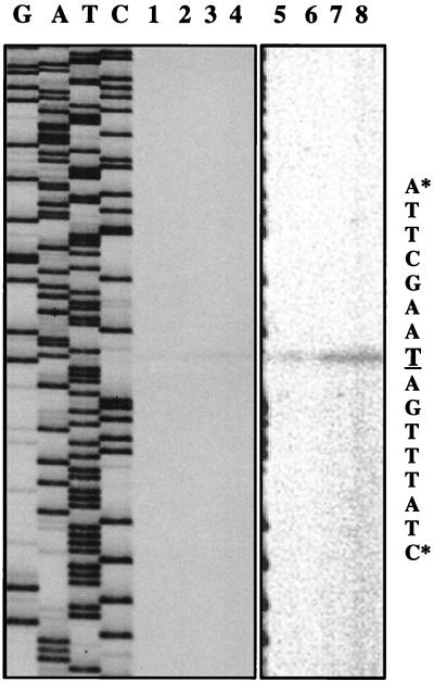
FIG. 4.
Primer extension analysis of the purR transcriptional start site. An autoradiogram shows primer extension experiments performed with 10 μg of RNA extracted from MK219 (MG1363 purR-lacLM, lanes 1 and 2) and MK221 (MK177 purR-lacLM, lanes 3 and 4). RNA was extracted from cells growing exponentially in GSA medium (lanes 1 and 3) or in the same medium supplemented with purines (lanes 2 and 4). Lanes G, A, T, and C, sequencing reactions. Asterisks indicate the limits of the nucleotide sequence, shown on the right, with the transcriptional start site underlined. The picture was scanned at 400 dpi with a Scan Jet 4c/T (Hewlett-Packard Co.) and DeskScan II version 2.3 software. The TIF file was imported into Top Draw version 3.1 for the addition of text. Lanes 5, 6, 7, and 8 are identical to lanes 1, 2, 3, and 4, respectively, except that the image was acquired with a Packard Instant Imager. The Instant Imager measures the radioactivity over the surface of the gel and is more sensitive than autoradiography.

