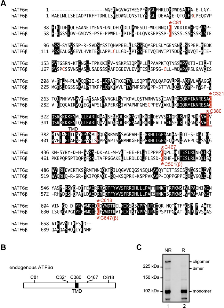
Fig. 1.
Amino acid alignment of human ATF6α and ATF6β. (A) ATF6α and ATF6β contain transmembrane domains (TMD, red square) in their middle. Amino acids identical between ATF6α and ATF6β are marked by white letters in black boxes. Five conserved cysteine residues are highlighted by white letters in red boxes with the asterisk and amino acid number. Two cysteine residues in the luminal region (C467 and C618 of ATF6α, and C501 and C647 of ATF6β) are highly conserved among vertebrates. (B) Schematic structure of ATF6α with the positions of TMD and five cysteine residues. (C) Immunoblotting using anti-ATF6α antibody of cell lysates prepared from WT HCT116 cells and then subjected to reducing (R) and non-reducing (NR) SDS-PAGE.

