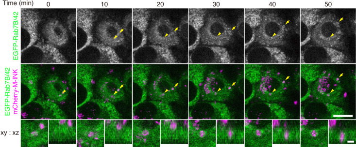
Fig. 4.
EGFP-Rab7B/42 accumulates around melanosomes incorporated into keratinocytes. Live imaging of EGFP-Rab7B/42 stably expressing in XB2 cells after adding mCherry-M-INK-labeled melanosomes. The images were stacked by z-projection. The arrowheads and arrows point to EGFP-Rab7B/42 accumulations around incorporated melanosomes (see also Supplemental Movies 1 and 2). Magnified views are stacked images of the incorporated melanosomes pointed to the arrowheads. Scale bars=15 μm (2 μm in magnified views).

