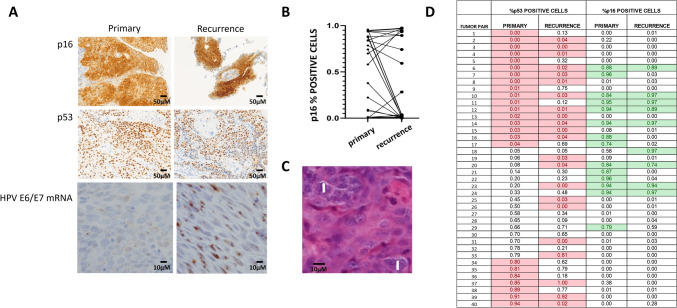
Fig. 1.
A Representative staining of primary and recurrent tumors for p16, p53 and high risk HPV E6/E7 mRNA. B Changes in p16 status between primary and recurrent tumors. C Examples of multinucleated cells denoted by white vertical bars within a primary tumor. D Shifts in p53 and p16 positivity expressed as fraction of cells noted to be positive in each primary or recurrent tumor sample from 40 tumor pairs. Red highlighted boxes denote aberrant p53 staining; green highlighted boxes denote p16 positivity in excess of 0.70 (scale bars represent 50µm and 10 µm, respectively)

