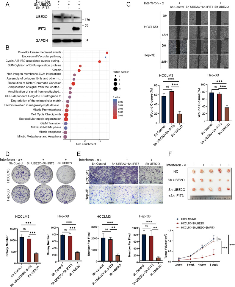
Fig. 6. IFIT3 reverses UBE2O knockdown-mediated interferon-α treatment potentiation.
Three kinds of cells were generated by transduction with the control, Sh-UBE2O, and Sh-IFIT3 lentiviral vectors, as shown in Fig. 6A. A Validation of the knockdown efficiency in the stably transduced cell lines. B Proteomic analysis was performed in the control and Sh-UBE2O+Sh-IFIT3 groups. The results showed that UBE2O no longer negatively regulated the interferon pathway after knockdown of IFIT3. Wound healing assays (C) colony formation assays (D) and migration assays (E) were used to verify the effect of the knockdown of UBE2O alone and simultaneous knockdown of UBE2O and IFIT3 on interferon sensitivity. Each set of experiments was performed with the same interferon concentration. The data are expressed as the mean ± SD of three independent experiments. *p < 0.05, **p < 0.01, *** p < 0.001, ns, nonsignificant. Ordinary one-way ANOVA with multiple comparisons testing was used to examine the statistical significance of differences among the three independent groups. F Stably transfected HCCLM3 cells from the control group, the UBE2O knockdown group, and the UBE2O and IFIT3 simultaneous knockdown group were injected into the inguinal region of mice, which were treated with the same dose of interferon, and the tumor volumes were calculated periodically (n = 5 mice/group). Ordinary one-way ANOVA with multiple comparisons testing was used for statistical analysis. ***p < 0.001, ns, nonsignificant.

