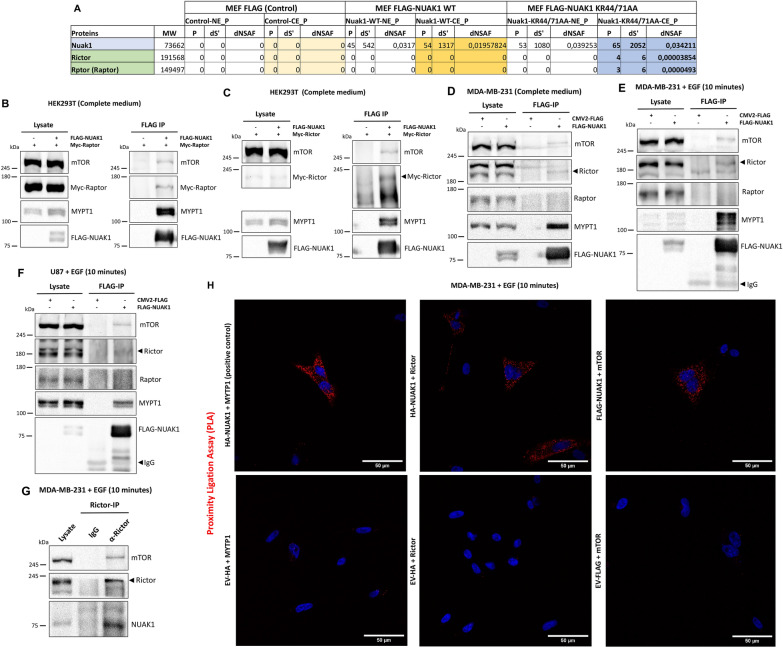
Fig. 1.
NUAK1 interacts with mTOR and Rictor but not with Raptor upon EGF stimulation. A Table shows total peptide count (P), the distributed spectra count (dS), and distributed normalized spectral abundance (dNSAF), observed for each identified protein in murine FLAG-NUAK1 WT and FLAG-NUAK1 KR44/71AA purifications (n = 3). NE, nuclear extract; CE, cytoplasmic extract. B Immunoblot (IB) of the Immunoprecipitation (IP) of human FLAG-NUAK1 WT and CoIP of endogenous mTOR, MYPT1 and exogenous Myc-Raptor in HEK293T cells. C IB of the IP of FLAG-NUAK1 WT and CoIP of endogenous mTOR, MYPT1 and exogenous Myc-Rictor in HEK293T cells. D IB of the IP of FLAG-NUAK1 WT and CoIP of endogenous mTOR, Rictor, Raptor and MYPT1 in MDA-MB-231 cells. E, F IB of the IP of FLAG-NUAK1 WT and CoIP of endogenous mTOR, Rictor, Raptor and MYPT1 from MDA-MB-231 (E) and U87 (F) cells serum-starved overnight before stimulation with EGF by 10 min. G IB of the IP of endogenous Rictor and CoIP of endogenous mTOR, and NUAK1 in MDA-MB-231 cells serum-starved overnight before stimulation with EGF by 10 min. H Proximity ligation assay (PLA) in MDA-MB-231 cells expressing HA-tagged NUAK1, FLAG-tagged NUAK1 or Empty vector (EV) (used as a negative control). Cells were serum-starved overnight and stimulated with EGF by 10 min (n = 3). Red dots indicate proximity of HA-NUAK1 with MYPT1 (Positive control), HA-NUAK1 with Rictor or FLAG-NUAK1 with mTOR. DAPI was used as a nuclear counterstain

