Figure 4.
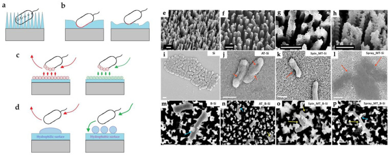
General strategies for the development of antibacterial surfaces are based on (a) contact killing, (b) the area of the contact zones, (c) electrostatic, and (d) wetting of the surface; (e–h) 45° view SEM images: (e) Black Si (B-Si) and (f) ALD-coated B-Si (AT_B-Si); mesoporous nanoparticles: (g) spin-coated (Spin_MT_B-Si) and (h) spray-coated (Spray_MT_B-Si) on B-Si (Scale bar: 1 μm). (i–p) Scanning electron microscopy images of bacteria on different substrates: (i) undamaged bacteria on flat Si; (j–l) bacterial cell wall disruption on ALD-, spin-, and spray-coated TiO2 surfaces, respectively; (m) bacterial cell has been pierced by nanostructures of black Si; (n–p) bacteria have sunken inside the TiO2-coated pillars, indicating cell death (Scale bar: 1 μm). Blue arrows indicate the piercing of bacteria by pillars. Red arrows indicate cell wall damaged areas. Yellow arrows indicate sunken bacteria. Adapted with permission from Ref. [47].

