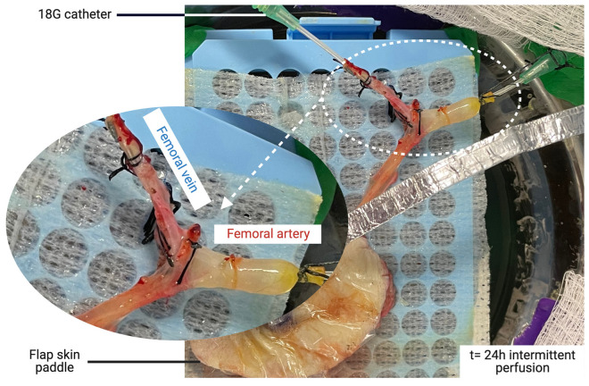
Figure 4.
Macroscopic aspect of the cannulated femoral vessels following 24 h of intermittent perfusion. The vein was cannulated to facilitate the procurement of the outflow sample from both veina comitans. Note that the positioning of the femoral vessels was adjusted while monitoring the system’s pressure to allow for perfusion with minimal mechanical resistance.

