Figure 3.
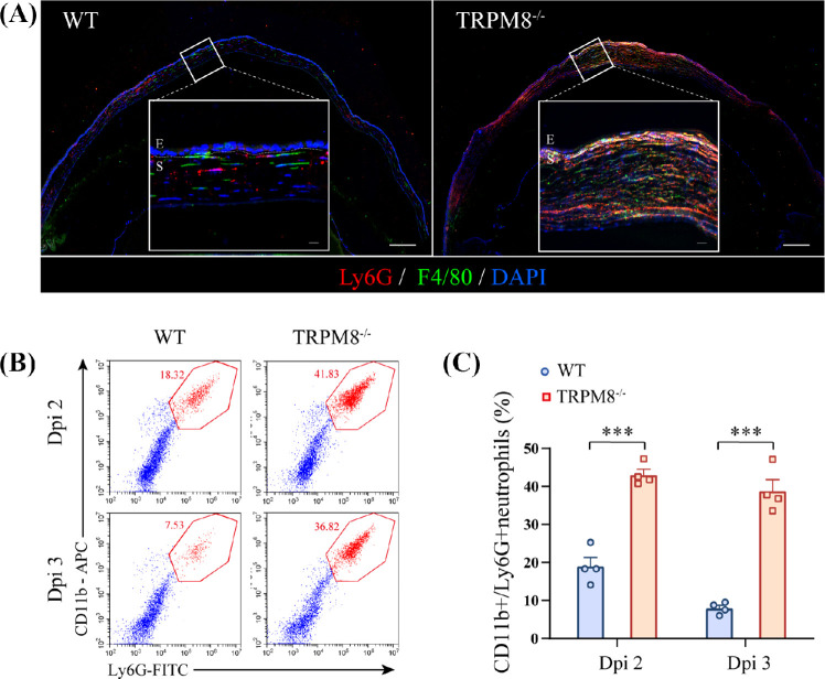
Recruitment and high persistence of CD11b+ Ly6G+ cells in TRPM8−/− mice in HSK. (A) Immunofluorescence for F4/80+ macrophages (green), Ly6G+ cells (red), and DAPI (blue) (Scale bar: 150 µm; E, epithelium; S, stroma) at dpi 3. White outlined insets show high-magnification images of the indicated regions (Scale bar: 25 µm; E, epithelium; S, stroma). Images were obtained from at least one of three independent experiments. (B) Representative dot plots of the percentage of CD11b+ Ly6G+ cells in the cornea analyzed 2 dpi and 3 dpi by flow cytometry in WT mice and TRPM8−/− mice. (C) Percentage of CD11b+ Ly6G+ cells in the cornea in WT mice and TRPM8−/− mice (n = 4 mice per group). Data are the mean ± SEM. ***P < 0.001.

