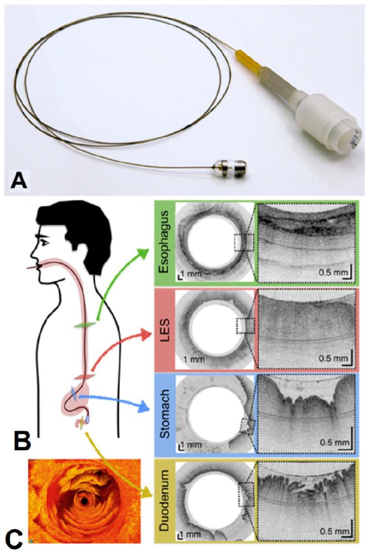
Figure 9.
(A) Photograph of the TCE. (B) A representation of the procedure and TEC images of the esophagus (green), lower esophageal sphincter (red), stomach (blue), and duodenum (gold). (C) Three-dimensional reconstruction of optical coherence tomography-based tethered capsule endomicroscopy (TCE) images, view of the second portion of the duodenum from a 30-year-old male participant [135]. Dashed lines show zooms on selected regions of TCE images.

