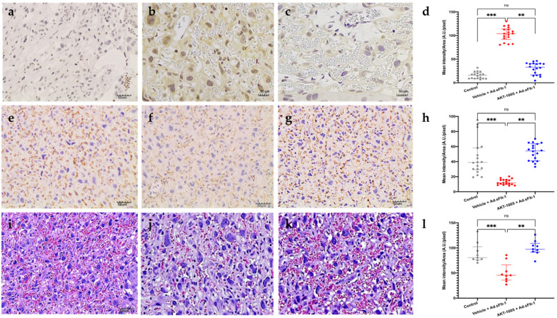
Figure 4.
Effect of AKT-1005 on placental stress in CD1 mice overexpressing sFlt-1. (a–d) Nitrotyrosine staining in placenta of pregnant mice given Ad.sFlt-1 on gd8 (induction of disease) and administered either AKT-1005 (20 mg/kg/day, (c)) or vehicle (b), starting on gd11 up to gd17 ((a), control pregnancy). Nitrotyrosine staining (indicative of nitrosative stress and peroxynitrite production) is significantly lower in the AKT-1005-treated group (c) compared with vehicle (b). (d) Quantitation of nitrotyrosine immunostaining: mean optical density was calculated in 6 high-power fields per sample (n = 3 per group). Kruskal–Wallis test, Dunn’s post hoc test, median (IQR). Control vs. Vehicle + Ad.sFlt-1: ***: p < 0.001, vehicle + Ad.sFlt-1 vs. AKT-1005 + Ad.sFlt-1: **: p < 0.01. (e–h) At gd17, AKT-1005 treatment improves CD31 staining abundance in the placenta (g) compared to vehicle-treated mice (f) ((e), control pregnancy). (h) Quantitation of CD31 immunostaining: mean optical density was calculated in 6 high-power fields per sample (n = 3 per group). Kruskal–Wallis test, Dunn’s post hoc test, median (IQR), control vs. vehicle + Ad.sFlt-1: ***: p < 0.001, vehicle + Ad.sFlt-1 vs. AKT-1005 + Ad.sFlt-1: **: p < 0.01. (i–l) At gd17, labyrinthine vasculature appeared collapsed in saline-treated mice (j). AKT-1005 improves placental vasculature in mice overexpressing sFlt1 (k). ((i), control pregnancy). (Original magnification: 20x, bar = 50 μm). (l) Quantitation of placental tissue vascular space: mean optical density was calculated in 3 high-power fields per sample (n = 3 per group). Kruskal–Wallis test, Dunn’s post hoc test, median (IQR), control vs. vehicle + Ad.sFlt-1: ***: p < 0.001, vehicle + Ad.sFlt-1 vs. AKT-1005 + Ad.sFlt-1: **: p < 0.01.

