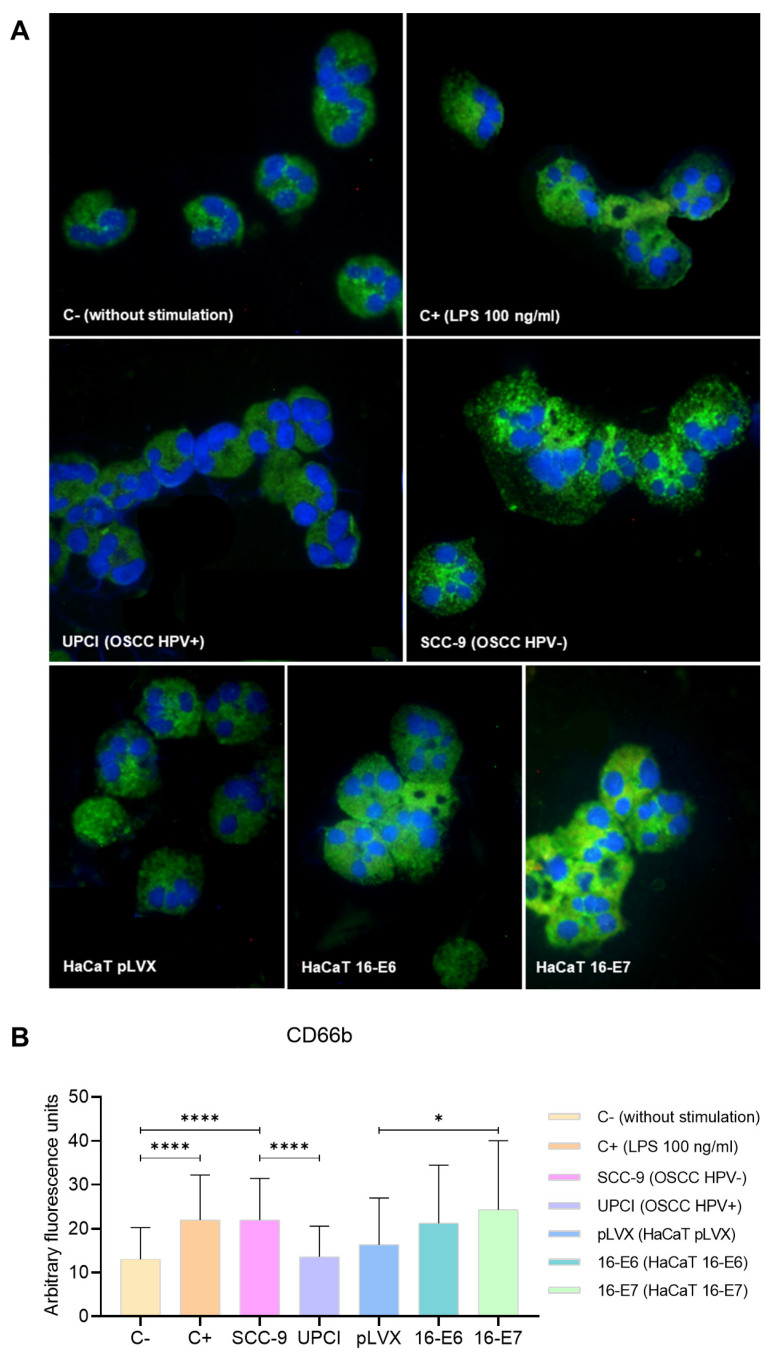
Figure 4.
SCC-9 (HPV−) cells induced overexpression of CD66b in neutrophils. CD66b expression was evaluated through immunofluorescence using an FITC-conjugated secondary antibody (green) and nuclear staining with DAPI (blue). Merged images are shown at 40× magnification (A), and the quantification is presented (B). Differences were found between C− and the other groups, except for HaCaT pLVX and UPCI:SCC154 (HPV+). Three independent experiments were performed. Data are presented as mean ± SD. * p < 0.05, **** p < 0.0001; one-way ANOVA, post hoc test: Games–Howell. 16-E6: HaCaT keratinocytes transduced with HPV16 E6, 16-E7: HaCaT keratinocytes transduced with HPV16 E7, C−: negative control, C+: positive control, LPS: lipopolysaccharide, pLVX: HaCaT keratinocytes transduced with the empty pLVX-Puro vector, SCC-9: HPV− OSCC cell line, UPCI: HPV+ OSCC cell line.

