Figure 1.
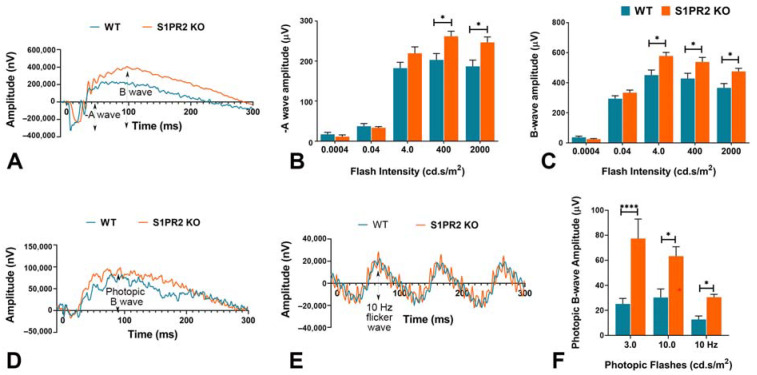
S1PR2 knockout (KO) mice exhibited higher baseline ERG responses than wild-type (WT) mice at 6 months of age. Shown are statistical analyses of scotopic ERG a- and b-wave amplitudes and photopic b-wave at different flash intensities for 6-month-old WT (blue) and S1PR2 KO (orange) mice. (A) Scotopic ERG amplitude waveforms (0 to 300 ms) of WT and S1PR2 KO mice. (B) At higher flash intensities (400 and 2000 cd.s/m2), S1PR2 KO mice demonstrated significantly higher mean scotopic a-wave ERG responses than WT mice. (C) S1PR2 KO mice demonstrated significantly higher mean scotopic b-wave ERG responses than WT mice at 4.0, 400, and 2000 cd.s/m2 flash intensities. (D) Photopic ERG amplitude waveforms (0 to 300 ms) of WT and S1PR2 KO mice. (E) ERG response amplitude waveforms elicited at 10 Hz flash flicker stimulus in WT and S1PR2 KO mice. (F) S1PR2 KO mice displayed significantly higher mean photopic b-wave ERG response amplitudes than WT mice at 3.0 cd.s/m2, 10.0 cd.s/m2, and 10 Hz flash flickering stimulus. Shown are mean values ± SEM. * p < 0.05, **** p < 0.0001 indicate significant differences between groups of WT and S1PR2 KO mice (n = 8 mice per genotype). Abbreviations used: ERG, electroretinography; KO, knockout; WT, wild-type.

