Figure 9.
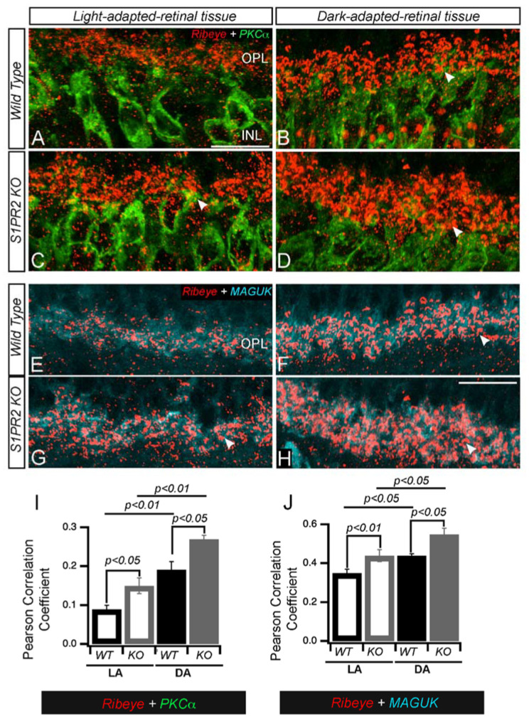
The retinas of S1PR2 KO mice exhibited altered photoreceptor ribbon synapses with post-synaptic densities (PSDs) in INL dendrites. (A–D) Transverse cross-sections of light-adapted (left panels (A,C,E,G)) or dark-adapted (right panels (B,D,F,H)) retinas from WT (A,B,E,F) or S1PR2 KO mice (C,D,G,H) were double immunostained with fluorescently labeled antibodies specific for either (A–D) the ribbon-specific marker ribeye (red) and the rod bipolar cell-specific marker PKCα (green) or (E–H) ribeye (red) and the post-synaptic marker MAGUK (cyan). Maximal intensity projections are shown; arrowheads indicate potential areas of colocalization. Scale bar, 50 µm. Abbreviations used: KO, knockout; MAGUK, membrane-associated guanylate kinase; PKCα, protein kinase C alpha; PSD, post-synaptic densities; WT, wild-type. (I,J) Pearson correlation analyses of IHC for accumulation of PKCα and ribeye or ribeye and MAGUK proteins in the OPL in LA and DA state represent the average value of the area of expression in 4–6 animals sampled per condition in 3–5 independent experiments. Statistical relevance was determined using a two-tailed t-test. Abbreviations used: IHC, immunohistochemistry; KO, knockout; OPL, outer plexiform layer; WT, wild-type; LA, light-adapted; DA, dark-adapted.

