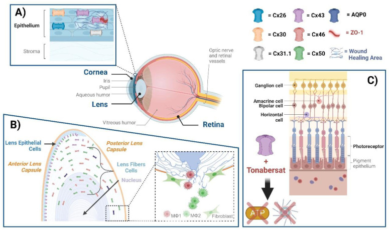
Figure 1.
Connexin GJs and HCs in eye diseases. The eye comprises the cornea, lens, retina, and several other subcomponents, within which various connexins exist. (A) Cx26, Cx30, Cx31.1, and Cx43 mediate GJ communication in the human corneal epithelium. ZO-1 is a scaffolding protein that plays a role in regulating GJ assembly. A disruption of Cx43 interaction with ZO-1 leads to the enhancement of GJs; targeting this disruption could be a means of enhancing wound healing. (B) Cx50 and AQP0, located at lens fibers, respectively, mediate cell–cell adhesion, maintaining lens fiber integrity. Deficiency of Cx50 and AQP0 leads to a loss in cell–cell adhesion, resulting in alterations of lens structures. At the posterior part of the lens, newly formed fibers cannot bundle properly, leading to lens posterior extrusion and capsule rupture. Macrophages are recruited to the vitreous cavity adjacent to the ruptured posterior capsule. M1 macrophages mainly mediate the removal of the tissue mass, while M2 macrophages play a crucial role in posterior capsule sealing and fibrosis. (C) The neuronal types of the retina are composed of photoreceptors, horizontal cells, bipolar, amacrine, and ganglion cells, all of which contain connexins. The participation of neuronal retinal Cxs in wound healing and fibrosis has not been directly reported. Tonabersat, a sodium channel blocker inhibiting Cx43 HCs, is used in some inflammatory-related retinal diseases. Tonabersat has also been shown to prevent the formation of NOD-like receptor protein 3 (NLRP3), an ATP-mediated inflammasome, as well as the release of other proinflammatory cytokines. Throughout the lens tissue, targeting Cxs for inflammation or fibrosis may hold therapeutic potential.

