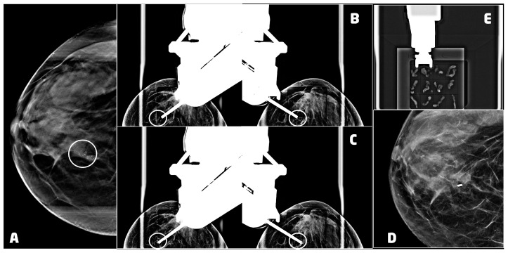
Figure 3.
VABB upright procedure for a 53 years old women with a ductal carcinoma in situ, G3 of the right breast: (A) digital breast tomosynthesis of the right breast, in cranio-caudal projection, showing a cluster of microcalcifications at the union of the inner quadrants (circle); (B) pre-fire stereotactic images of the right breast showing the position of the needle near the suspicious lesion (circle); (C) post-fire stereotactic images of the right breast showing the position of the needle inside the suspicious lesion (circle); (D) 2D synthetic view of the right breast 2 days after the procedure, showing the 3mm titanium clip marking the biopsy site with neither residual microcalcifications nor hematoma; (E) specimen radiograph showing microcalcifications in the samples.

