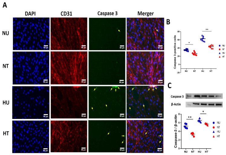
Figure 4.
LA shields against apoptosis in PBECs under hypoxic and hyperglycemic conditions, 24 h after LA treatment. (A) Representative fluorescent microscopic images of triple staining of PBECs for apoptotic marker, casepase-3 marker; DAPI (blue), CD-31 (red), cleaved caspase-3 (green); the merger. More intense cleaved caspase-3 signals were counted for quantification (pointed yellow arrows) in all four groups of cells. (B) Quantification of the number of cleaved caspase-3-positive cells 24 h after LA treatment under hypoxic and glycemic conditions (hyper- and/or normoglycemic). LA-treated NT cells show significantly fewer cleaved caspase-3 fluorescent signals than HU cells. (C) Quantification of immunoblotting for cleaved caspase-3 in four groups of PBECs: HU, HT, NU, and NT. LA reduced phosphorylation of apoptotic proteins in the presence of LA in hyperglycemic and normoglycemic groups compared to untreated cells. Analysis followed a two-way ANOVA with Bonferroni’s post hoc test. (B,C) Scale bar is 50 µm. Values shown as means ± SD; * p < 0.05; ** p < 0.01.

