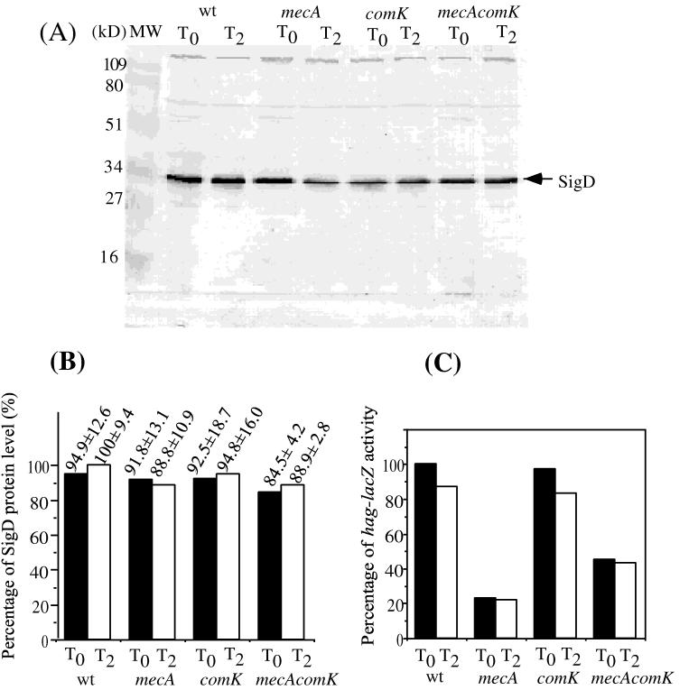
FIG. 3.
Level of ςD protein in the wild type (wt) and mecA, comK, and mecA comK mutants grown in rich medium. Cells precultured in 2XYT liquid medium were grown in 2XYT medium at 37°C. Samples were harvested at T0 and T2 (at the end of exponential growth and 2 h after the end of exponential growth, respectively). Cell extracts with equal protein concentrations were applied to SDS–12% polyacrylamide gels and subjected to electrophoresis. The resolved protein was electrotransferred to nitrocellulose and analyzed by the Western blotting procedures described in Materials and Methods. (A). Lane MW contained molecular size markers. The other lanes contained samples from cultures of LAB2607 (wild type), LAB2722 (mecA), LAB2723 (comK), and LAB2724 (mecA comK) cells collected at T0 and T2 of the growth curve. (B) Western blot band intensity determined by scanning of the image of the stained blot and quantification by the NIH-Image computer program. The values presented are percentages of the level of protein in the wild-type strain at T2. The standard deviation was calculated from three independent experiments. (C) Levels of hag-directed β-galactosidase in cultures used to obtain extracts for Western blot analysis.

