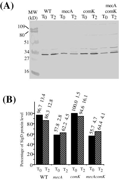
FIG. 4.
Levels of ςD protein in the wild type (WT) and mecA, comK, and mecA comK mutants grown in CM. Cells precultured in DSM agar plates were grown in CM at 37°C. Samples were harvested at the same time points as in Fig. 3. Analysis of the protein extracts was conducted as described in the legend to Fig. 3. (A) Western blot of extracts of LAB2607 (wild type [WT]), LAB2722 (mecA), LAB2723 (comK), and LAB2724 (mecA comK) cell samples collected at T0 and T2 of the growth curve. (B) Western blot band intensity determined and presented as described in the legend to Fig. 3.

