Figure 3.
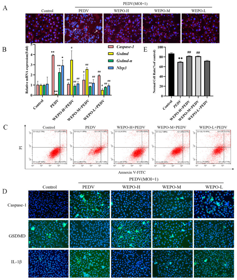
GSDMD participates in the pyroptosis of WEPO-inhibited PEDV-infected Vero cells. (A) The integrity of the cell membrane was examined by Dil staining after infection with PEDV and the addition of different concentrations of WEPO. (The magnification of a microscope is 200 times. The white arrow indicates the place where the Vero cells have lesions and rupture, and the cell membrane fuses). (B) RT-PCR was used to detect the expression of Caspase-1, GSDMD, GSDMD-N, and Nlrp3 in Vero cells after infection with PEDV and the addition of different concentrations of WEPO. The data are expressed as the means ± SD (n = 3). * p < 0.05 and ** p < 0.01 vs. control. # p < 0.05 and ## p < 0.01 vs. positive control (PEDV). (C) Detection of the degree of apoptosis in Vero cells by flow cytometry. (D) The expression of Caspase-1, GSDMD, and IL-1β in Vero cells was detected by immunofluorescence after infection with PEDV and treatment with different concentrations of WEPO. (The magnification of a microscope is 200 times). (E) Flow cytometry data analysis histogram. The data are expressed as the means ± SD (n = 3). ** p < 0.01 vs. control. ## p < 0.01 vs. positive control (PEDV).

